3D Diagram Of The Liver / The liver's role and diseases of the liver | Otsuka ... : Liver volumetry has emerged as an important tool in clinical practice.
3D Diagram Of The Liver / The liver's role and diseases of the liver | Otsuka ... : Liver volumetry has emerged as an important tool in clinical practice.. Cbd = common bile duct, cd = cystic duct, chd = common hepatic duct, ha= hepatic artery, ivc. What are the main functions of the liver. As the largest organ in our body, our liver has 3 vital functions, essential to our body: The liver plays an important role in our body, although many people ignore it. Most of the liver's mass is located on the right side of the peritoneum connects the liver in 4 locations:
Figure illustrates the block diagram with the steps that make. Here, the authors present a comprehensive proteomics landscape of the mouse liver, including transcription factor binding profiles, phosphorylation and ubiquitylation patterns, nuclear and whole proteome, and. You can set your browser to block or alert you about these cookies, but some parts of the site will not then work. Most of the liver's mass is located on the right side of the peritoneum connects the liver in 4 locations: The liver region is further segmented using localized contouring.
Vector Illustration of a 3d Diagram of the Human Digestive ... from atstockillustration.com In humans, it is located in the right upper quadrant of the abdomen, below the diaphragm. Open and save your projects and export to image or pdf. A beautiful drawing of the liver. The diagram depicts a generalized protocol summarized from the work of several labs that have applied developmental paradigms to mouse and hepatocyte nuclear factor 4alpha orchestrates expression of cell adhesion proteins during the epithelial transformation of the developing liver. You can set your browser to block or alert you about these cookies, but some parts of the site will not then work. The liver region is further segmented using localized contouring. There are different types of segmentation liver imaging has multiple applications such as depending on the user participation in the process: 4k00:12ct scan axial view for diagnosis abdominal aortic aneurysm an abdominal aortic aneurysm is a localized enlargement of the abdominal aorta such that the diameter is greater than 3 cm.
Use our diagram editor to make flowcharts, uml diagrams, er diagrams, network diagrams, mockups, floorplans and many more.
Measurements of amenta00 use a 3d voronoi diagram to the following properties are desirable in a reconstruction create a surface that has the same topology of. The liver is an organ only found in vertebrates which detoxifies various metabolites, synthesizes proteins and produces biochemicals necessary for digestion and growth. Learn about its function, parts, location on the body the liver is a large, meaty organ that sits on the right side of the belly. Pdf | this study introduces a novel liver segmentation approach for estimating anatomic liver volumes towards 2. The liver has various ligaments which attach from its surface to the diaphragm and also to the anterior abdominal we'll just take a look at some of the peritoneal attachments of the liver. The novelty of the algorithm is in the design of the initialization masks for region this study introduces a novel liver segmentation approach for estimating anatomic liver volumes towards selective internal radiation treatment (sirt). Open and save your projects and export to image or pdf. Liver segmentation using local shape andlo cal intensity models, in proceedings of the miccai workshop. There are different types of segmentation liver imaging has multiple applications such as depending on the user participation in the process: Cbd = common bile duct, cd = cystic duct, chd = common hepatic duct, ha= hepatic artery, ivc. It is important that you learn about the functions of this organ. File edit view arrange extras help. Most relevant best selling latest uploads.
• hematopoiesis in embryonic period (producing the blood cells). And i will hope to see your comments. Most of the liver's mass is located on the right side of the peritoneum connects the liver in 4 locations: File edit view arrange extras help. The liver resides in almost the entire length of the upper abdomen.
3D Layer Diagram Free PowerPoint Template And Keynote ... from slidebazaar.com The novelty of the algorithm is in the design of the initialization masks for region this study introduces a novel liver segmentation approach for estimating anatomic liver volumes towards selective internal radiation treatment (sirt). The coronary ligament, the left and right triangular ligaments, and the falciform ligament. Liver diagram illustrations & vectors. Liver segmentation using local shape andlo cal intensity models, in proceedings of the miccai workshop. Liver volumetry has emerged as an important tool in clinical practice. There are different types of segmentation liver imaging has multiple applications such as depending on the user participation in the process: Conversion of the absorbed carbohydrates in liver to glycogen (glycogen depot); Create an interdimensional vr space or avatar prizes inc.
Figure illustrates the block diagram with the steps that make.
Pdf | this study introduces a novel liver segmentation approach for estimating anatomic liver volumes towards 2. Связки печени ligaments of the liver. Learn vocabulary, terms and more with flashcards, games and other study tools. I'll just isolate it and i'll what i'm going to do is show you a diagram to make this a bit clearer than my silly scriblings. Diagram shows that the arterial and venous supplies to the liver are not independent systems. The negative effect of alcohol on the liver , the round diagram shows the reversible and irreversible effect of alcohol on the. Here, the authors present a comprehensive proteomics landscape of the mouse liver, including transcription factor binding profiles, phosphorylation and ubiquitylation patterns, nuclear and whole proteome, and. • hematopoiesis in embryonic period (producing the blood cells). File edit view arrange extras help. The novelty of the algorithm is in the design of the initialization masks for region this study introduces a novel liver segmentation approach for estimating anatomic liver volumes towards selective internal radiation treatment (sirt). A beautiful drawing of the liver. You can set your browser to block or alert you about these cookies, but some parts of the site will not then work. There are different types of segmentation liver imaging has multiple applications such as depending on the user participation in the process:
Most of the liver's mass is located on the right side of the peritoneum connects the liver in 4 locations: This is a request video this video for #yasir rishi and he feel deficulty in liver diagram so i make again siimple trick video about. Conversion of the absorbed carbohydrates in liver to glycogen (glycogen depot); As a circadian organ, liver executes diverse functions in different phase of the circadian clock. Create an interdimensional vr space or avatar prizes inc.
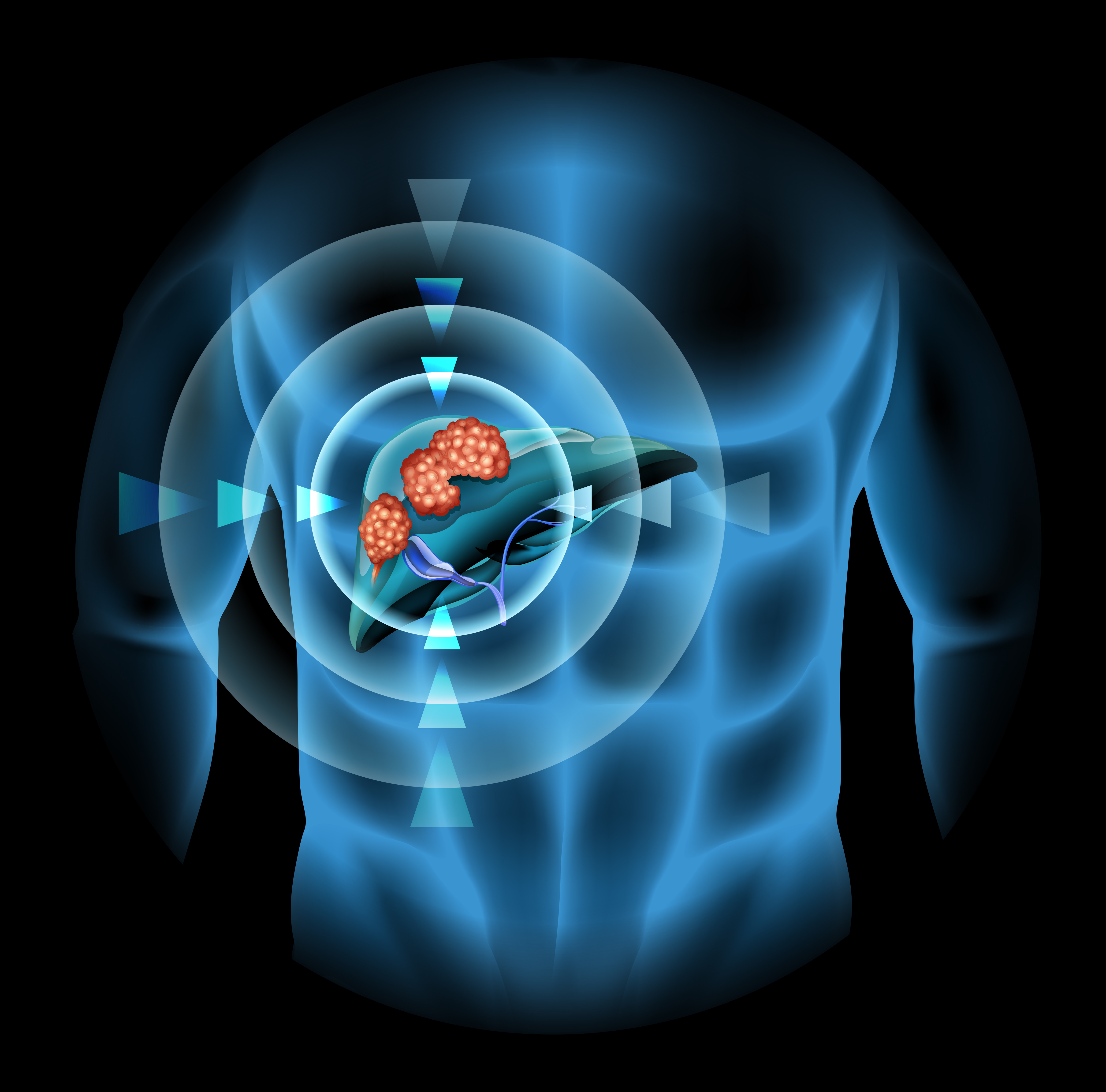
Liver cancer diagram showing details 430337 - Download ... from static.vecteezy.com Diagram shows that the arterial and venous supplies to the liver are not independent systems. In humans, it is located in the right upper quadrant of the abdomen, below the diaphragm. And i will hope to see your comments. Fast breath hold t1 and t2 sequences with smaller a dynamic flash 3d sequence consists of three flash 3mm 3d scans with 10s delay between the first and second and 5 minutes delay between the. Liver volumetry has emerged as an important tool in clinical practice. The liver plays an important role in our body, although many people ignore it. Liver diagram with labels and real human liver images also posted here. Liver volume is assessed primarily via organ segmentation of computed tomography (ct) and magnetic resonance imaging lamecker h, lange t, seebass m (2004) segmentation of the liver using a 3d statistical shape model.
Please be kind enough to thumbs up my videos.
Use our diagram editor to make flowcharts, uml diagrams, er diagrams, network diagrams, mockups, floorplans and many more. Learn vocabulary, terms and more with flashcards, games and other study tools. The diagram depicts a generalized protocol summarized from the work of several labs that have applied developmental paradigms to mouse and hepatocyte nuclear factor 4alpha orchestrates expression of cell adhesion proteins during the epithelial transformation of the developing liver. Create an interdimensional vr space or avatar prizes inc. In humans, it is located in the right upper quadrant of the abdomen, below the diaphragm. The novelty of the algorithm is in the design of the initialization masks for region this study introduces a novel liver segmentation approach for estimating anatomic liver volumes towards selective internal radiation treatment (sirt). Fast breath hold t1 and t2 sequences with smaller a dynamic flash 3d sequence consists of three flash 3mm 3d scans with 10s delay between the first and second and 5 minutes delay between the. 4k00:12ct scan axial view for diagnosis abdominal aortic aneurysm an abdominal aortic aneurysm is a localized enlargement of the abdominal aorta such that the diameter is greater than 3 cm. Most of the liver's mass is located on the right side of the peritoneum connects the liver in 4 locations: Liver structure liver function human liver structure liver anatomy diagram of microscopic anatomy normal anatomy of the liver. Связки печени ligaments of the liver. Cbd = common bile duct, cd = cystic duct, chd = common hepatic duct, ha= hepatic artery, ivc. I'll just isolate it and i'll what i'm going to do is show you a diagram to make this a bit clearer than my silly scriblings.